
Figure 4.
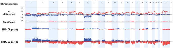
Cumulative chromosomal aberrations of 22 infant cases in comparison to 16 non‐infant pediatric high‐grade gliomas. Molecular inversion probe (MIP) analysis shows that chromosomal alterations are significantly less frequent in diffuse infant high‐grade gliomas (iHGGs) compared to pediatric high‐grade gliomas (pHGGs). The comparison plot is obtained by subtracting the genomic alterations of groups. The difference is expressed in percentage for the gains and losses for each location (gain and loss are shown as up and down, respectively) and tracked in the upper part of the figure. Bars in the track “Significant” indicate regions where there is a significant difference of the copy number changes between the compared groups (P < 0.05). The profile of copy number changes (losses in red, gains in blue) for each group is displayed in the lower track of the plot.