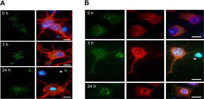
Figure 2.

The distribution of calsenilin and presenilin (PS) 1 in primary cortical neurons subjected to ischemia‐like conditions. A. Calsenilin immunofluorescence in mouse primary cortical neurons, showing calsenilin (green), the neuronal marker MAP2 (red) and 4’,6‐diamidino‐2‐phenylindol (DAPI) (blue). Calsenilin staining showed a small, non‐significant increase after 3 h of glucose deprivation (GD). At 24 h, calsenilin congregated at nuclear peripheries. Staining was prominent around apoptotic bodies (asterisk). B. Calsenilin (green) and PS1 (red) immunofluorescence in mouse primary cortical neurons, both showing a small increase with GD. Calsenilin, but not PS1, congregated around apoptotic bodies (GD for 3 h, asterisk). At 24 h, calsenilin and PS1 signals co‐localized around nucleus. Scale bar: 10 μm.
