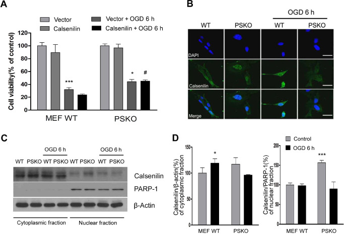
Figure 5.

Calsenilin over‐expression in PS‐1, 2 double knockout (PSKO) cells reduces OGD‐induced cell death. A. No significant cell death rate difference was observed between WT and PSKO mouse embryonic fibroblast (MEFs) subjected to OGD. However, after cells had been transfected with calsenilin, PSKO MEFs showed significantly lower death rates than WT MEFs. Cell viability determined by luminescent cell viability assay. Values are the mean with standard error of measurement (SEM). (n = 4). *P < 0.05, ***P < 0.001 vs. vector + control, # P < 0.05 MEF WT calsenilin + OGD6h. B. PSKO MEFs, exposed to OGD for 6 h and then immunostained with anti‐calsenilin, showed a cell‐wide calsenilin distribution whereas in WT MEFs, calsenilin was restricted to the nucleus. Original magnification, 1000×. Scale bar: 50 μm. C. Representative immunoblot of cytoplasmic and nuclear fractions of WT and PSKO MEF cells showed that nuclear localization of calsenilin was significantly increased in PSKO MEF cells compared with WT MEF cells, however nuclear calsenilin of PSKO MEF cells decreased significantly after OGD. D. Quantification of calsenilin levels normalized to a loading control (β‐actin for cytoplasmic fraction; Poly [ADP‐ribose] polymerase‐1 (PARP‐1) for nuclear fraction). Values are the mean with SEM. (n = 3) *P < 0.01, ***P < 0.001 vs. MEF WT + control.
