
Figure 1.
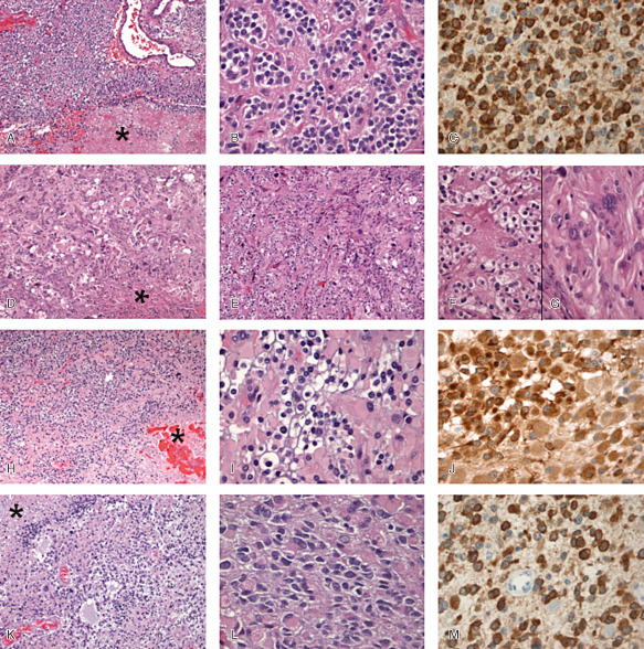
A–C. Anaplastic oligodendroglioma (AO) with focal necrosis. A. Oligodendroglial population with high cellularity, vascular proliferation and focal necrosis (asterisk) [hematoxylin and eosin (HE) ×100]. B. Perinuclear halo, moderate pleomorphism and hyperchromatism (HE ×400). C. Diffuse expression of IDH1 R132H (×400). D–G. Glioblastoma with oligodendroglial component (GBM/GBMO). D. Malignant glioma with high cellular pleomorphism and necrosis (asterisk) (HE ×100). E. On the left, tumor is composed of round cells with perinuclear halo and on the right by more pleomorphic cells (HE ×100). F. High magnification of oligodendroglial‐like component (HE ×400). G. High magnification of more classical GBM feature (HE ×400). H–J. Anaplastic oligoastrocytoma (AOA) with necrosis/GBMO. H. Mixture of oligodendroglial‐like and gemistocytic‐like cells with necrotic area (asterisk) (HE ×100). I. High magnification showing transitional appearances between oligodendroglial and astrocytic cells (HE ×400). J. Diffuse expression of IDH1 R132H (×400). K–M. AOA with necrosis/GBMO. K, L. Mixture of oligodendroglial cells and astrocytic cells usually pleomorphic with necrotic area (asterisk) (HE ×100). M. Diffuse expression of IDH1 R132H (×400).