
Figure 3.
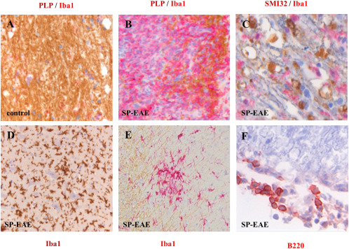
Histopathology of secondary progressive EAE in Biozzi ABH mice. Compared to aged‐matched controls in which myelin integrity is intact and in which few if any Iba‐1+ cells are present (A), in SP‐EAE demyelination is demarcated by the loss of PLP associated with highly immunereactive Iba‐1+ microglia/macrophages (B). In white matter, SMI32 depicting damaged axons is prominent in regions of highly reactive Iba‐1+ cells (C). In addition to Iba‐1 positive cells in the white matter, large infiltrates are also observed in gray matter in close association with motor neurons (D). Like MS, clusters of activated microglia are observed in normal appearing white matter in mice with SP‐EAE (E). In comparison to acute EAE in Biozzi ABH mice, a sparse number of adaptive immune cells are observed during SP‐EAE, and when observed are generally restricted to the leptomeninges (F).