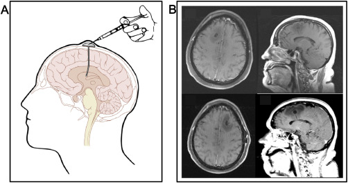
Figure 4.

Advances in Ommaya reservoirs. A. Schematic representation of an implanted Ommaya reservoir. After an artwork by Patrick J. Lynch, with permission. B. MRI of the brain of a 65‐year old woman with an implanted Ommaya reservoir for intraventricular therapy with topotecan, showing a cystic dilatation of the brain parenchyma with vasogenic edema surrounding the catheter connecting to the reservoir dome and delivering the drug to the intraventricular space (Figure 4B, first row). After removing the Ommaya reservoir because of neurological symptoms including left‐sided weakness, confusion and headaches, the neurosurgeons found no evidence of tumoral infiltration or infection and another Ommaya reservoir was placed in the contralateral side in order to avoid discontinuation of therapy. After 7 treatments, the patient displayed yet similar neurological symptoms and radiological findings and the treatment had to be halted (Figure 4B, second row) (after Ref. 35 with permission).
