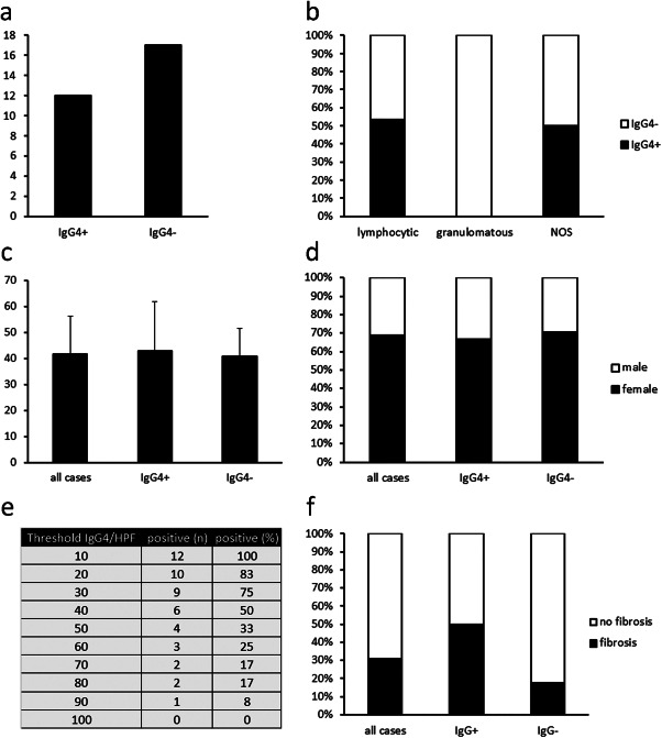
Figure 2.

Statistical analysis of hypophysitis cases. (a) Absolute numbers of cases previously diagnosed as hypophysitis fulfilling the criteria for IgG4‐related hypophysitis (IgG4+) or failing to fulfill these criteria (IgG4−). (b) Percentages of IgG4+ and IgG4− hypophysitis among cases previously diagnosed as lymphocytic hypophysitis, granulomatous hypophysitis, or hypophysitis, not otherwise specified (NOS). (c) Mean ages ± standard deviation of all 29 cases analyzed and of IgG4+ and IgG4− hypophysitis. (d) Percentages of male and female patients among all cases and IgG4+ and IgG4− hypophysitis. (e) Sensitivity of different thresholds for the number of IgG4‐positive cells per high power field in the diagnosis of IgG4‐related hypophysitis. (f) Percentages of storiform fibrosis among all cases and IgG4+ and IgG4− hypophysitis.
