
Figure 2.
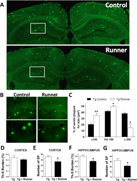
Running reduces the levels of Th‐S burden in APPswe/PS1ΔE9 mice brain. A. Amyloid plaques detected with Th‐S staining in control (Tg) and runner (Tg runner) APPswe/PS1ΔE9 mice brain. Images represent a reconstruction of the hippocampus and cortex of control and runner transgenic mice. B. Magnification of senile plaques. C. The graphs classified the senile plaques of control (Tg) and runner (Tg runner) APPswe/PS1ΔE9 mice for size (mm2). Amyloid burden was quantified with the Th‐S staining and the percentage of the area covered by amyloid plaques (D) and the number of senile plaques (E) in cortex and hippocampus, respectively (F,G), was plotted. Bars represent mean ± SE (n ≥ 5). *P < 0.05; **P < 0.01.