Figure 4.

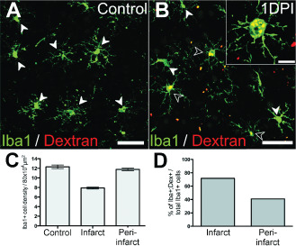
Infiltration of peripheral macrophages in the acute post‐ischemic period. Iba1+/dextran+ peripheral macrophages were detected in brain tissues proximal to lesion site at 1 days post ischemia (DPI) (B; enlarged in insert), but not in controls (A). White arrowheads denote Iba1+/dextran− macrophages. Empty arrowheads denote Iba1+/dextran+ macrophages. (C–D). Quantitative analysis of local and infiltrating macrophages at the infarct and peri‐infarct areas at 1 DPI. Total macrophage populations (Iba1+) was lower at the infarct site compared with peri‐infarct area (600–800 μm distal to core), which remained close to control levels. The percentage of infiltrating (Iba1+/dextran+) over total macrophage (Iba1+) population was greater in the infarct core compared with the peri‐infarct area. Scale: (A, B) 50 μm, (B insert) 20 μm.
