
Figure 1.
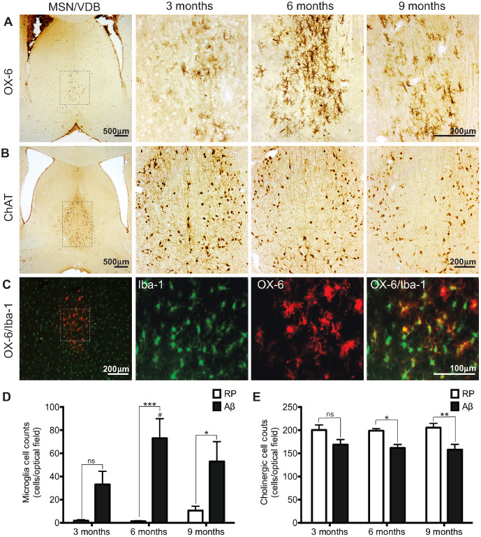
Basal forebrain pathology. Representative images of (A) OX‐6 immunopositive microglia and (B) choline acetyltransferase (ChAT) immunolabeled cholinergic neurons in the medial septal nucleus/vertical diagonal band (MSN/VDB) of the basal forebrain in Aβ25–35 administered animals 3, 6 and 9 months of age. C. Microglia in the MSN/VDB of a 6‐month‐old Aβ25–35 administered rat identified with two different microglia markers; OX‐6 and Iba‐1. All OX‐6 positive microglia co‐localized with Iba‐1 positive microglia. Boxed areas in low magnification images in panel 1 illustrate the location of high magnification pictures in panels 2–4. (D) The number of OX‐6 immunoreactive microglia and (E) the number of ChAT positive neurons in the MSN/VDB of Reverse peptide Aβ35–25 and Aβ25–35 administered rats, 3, 6 and 9 months of age. Data presented as mean ± SEM, n = 5–7 animals/group; *P < 0.05; **P < 0.01; ***P < 0.001; ns—not significant between treatment groups within an age group; #P < 0.05 6 months Aβ vs. 3 months Aβ.