
Figure 3.
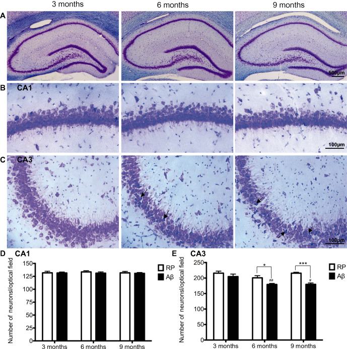
Hippocampal pathology. A–C. Representative photomicrographs of thionin stained CA1 and CA3 hippocampal subfields, in Aβ25–35 administered rats 3, 6 and 9 months of age. Arrows indicate representative misshapen/irregular neurons. D–E. Average numbers of hippocampal neuronal counts in the CA1 and CA3 subregions of the hippocampus, determined 3 weeks following intracerebroventricular administration of reverse peptide (RP) Aβ35‐25 or Aβ25–35 in 3, 6 and 9‐month‐old rats. Data presented as mean ± SEM, n = 6/7 animals/group; *P < 0.05; ***P < 0.001 between treatment groups within an age group; ##P < 0.01 6 months Aβ vs. 3 months Aβ; #P < 0.05 9 months Aβ vs. 3 months Aβ.