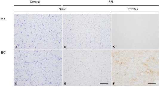
Figure 2.

Mediodorsal nucleus of the thalamus (thal: A–C) and entorhinal cortex (EC: D–F) in control (A,B) and FFI (B,C,E,F) cases showing marked loss of neurons without spongiform change in the thalamus and moderate neuron loss and spongiform change in entorhinal cortex in FFI when compared with corresponding regions in controls. In FFI, no proteinase K‐resistant PrP (PrPRes) is observed in the mediodorsal thalamus (C) whereas punctate, synaptic‐like immunostaining is observed in the entorhinal cortex (F) processed in parallel. Paraffin sections, A, B, D, E Nissl staining, bar in E = 50 µm; C, F, PrP immunohistochemistry (3F4 antibody) following preincubation with proteinase K, bar in F = 25 µm.
