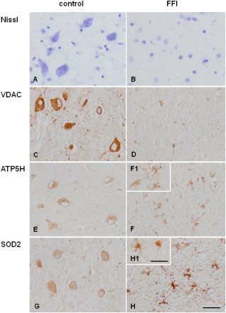
Figure 4.

Mediodorsal nucleus of the thalamus in control (A, C, E, F) and FFI (B, D, F, H) cases. Nissl staining (A, B) shows marked decrease in the number of neurons in FFI when compared with controls. (VDAC) (C, D), (C, D) (SOD2) (E, F) immunoreactivity is found in mediodorsal thalamus in control (A, C, E) and FFI (B, D, F) cases). Decreased VDAC (C, D) and ATP synthase, H+ transporting, mitochondrial Fo complex, subunit d (ATP5H) (E, F) immunoreactivity is found in the mediodorsal thalamus in FFI compared with controls due to the dramatic decrease in the number of neurons. However, ATP5H is observed in reactive glial cells. Weak superoxide dismutase 2 (SOD2) (G, H) immunoreactivity is seen in neurons in control cases which contrasts with enhanced SOD2 immunostaining in reactive astrocytes in FFI. Paraffin sections, A, B: Nissl staining; C–H immunohistochemical sections slightly counterstained with haematoxylin. A–H, bar in H = 25 µm. F1, H1, bar in H1 = 10 µm.
