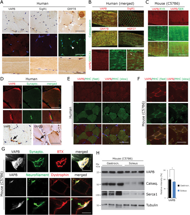
Figure 1.

VAPB expression profile in normal innervated human and mouse muscle fibres. A. Human muscle biopsy paraffin sections of normal human control muscles (n = 6, upper and middle row: transverse section, lower row: longitudinal sections) showing a pattern of VAPB, SigR1 and GRP78 immunoreactivity associated with sarcomeric bands after diaminobezidine and immunofluorescence labelling. Note the preferential GRP78 staining of the envelope of myonuclei (arrowheads). One myonucleus is shown at higher magnification in the inset. Scale bar = 40 µm. B. Merged immunofluorescence staining of VAPB, SigR1, GRP78 and HSP27 immunoreactivities associated with sarcomeric bands. Immunofluorescence double‐labelling demonstrates colabelling with calsequestrin (upper and middle panels) and with SERCA1 (lower panel) in normal‐sized, innervated fibres (human muscle biopsy paraffin sections). Scale bar = 30 µm. C. VAPB colocalization with RyR1 (left panel) and with SERCA1 (right panel) in normal‐sized, innervated mouse gastrocnemius muscle fibres. Scale bar = 25 µm. D. Immunofluorescence and DAB staining (lower panel) showing intense VAPB labelling of the postsynaptic sarcoplasm at human NMJs costained for the presynaptic marker synaptophysin. GRP78 immunoreactivity showed a similar pattern (human muscle biopsy, paraffin sections). Scale bar = 30 µm. E. VAPB expression does not differ between slow compared to fast muscle fibres in normal human muscle; immunofluorescence using antibodies against VAPB as well as fibre type‐specific myosin heavy chain isoforms (human muscle biopsy, paraffin sections). Scale bar = 50 µm. F. VAPB is expressed at slightly decreased levels in slow compared to fast muscle fibres in normal mouse muscle; immunofluorescence using antibodies against VAPB as well as fibre type‐specific myosin heavy chain isoforms. Scale bar = 50 µm. G. VAPB immunofluorescence of NMJs of teased normal mouse muscle fibres costained for the presynaptic marker synaptophysin and for the postsynaptic marker alpha‐bungarotoxin (BTX) shows prominent VAPB immunoreactivity. Lower panel: triple labelling with antibodies against VAPB, neurofilament protein and dystrophin shows postsynaptic localization of VAPB immunoreactivity. Scale bar = 40 µm. H. Western blot analysis of VAPB protein demonstrating significantly lower expression levels in soleus vs. gastrocnemius muscles in normal mice (n = 3). There are even lower levels of the Ca2+‐binding proteins calsequestrin and SERCA1 in soleus compared to gastrocnemius muscle. The graph illustrates the results of the quantitative densitometric analysis of the VAPB levels. *P < 0.05.
