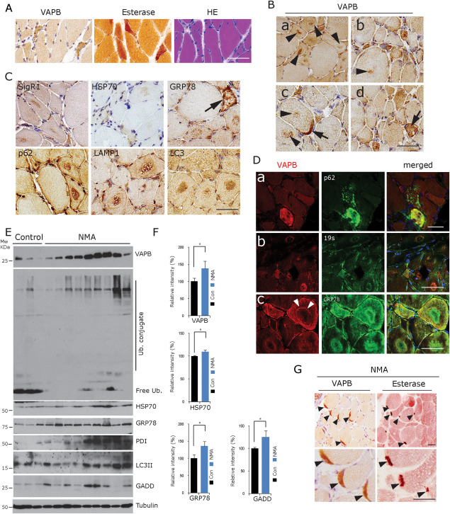
Figure 2.

VAPB accumulation in denervated fibres in NMA. A. Accumulation of VAPB in denervated, partially atrophic and atrophic human muscle fibres. They are angular or flattened, grouped and often esterase‐positive. H&E staining, esterase histochemistry and VAPB immunohistochemistry of consecutive human muscle biopsy frozen sections from a representative case from the group of 11 NMA cases available for cryostat sectioning. Scale bar = 100 µm. B. Prominent VAPB staining of target structures in NMA cases [arrowheads in (a–c)]. The centre of the targets and ring‐like structures around targets were VAPB immunoreactive. Strong immunostaining of VAPB at an NMJ [arrow in (c)] and of a muscle fibre showing secondary myopathic alterations [arrow in (d)] (human muscle biopsy, paraffin sections). Scale bar = 50 µm. C. Staining with antibodies against other ER chaperones (SigR1, HSP70 and GRP78) and against autophagy markers (p62, Lamp1 and LC3) reveals accumulations of these proteins in the centre of target structures. Arrow: strongly GRP78‐positive lesioned muscle fibre (human muscle biopsy, paraffin sections). Scale bar = 50 µm. D. Considerable immunofluorescence colabelling of VAPB with p62 (upper panel), 19s proteasome subunit (middle panel) and GRP78 (lower panel) in lesioned fibres, targets and atrophic muscle fibres in NMA. Note the prominent VAPB‐labelling of the rim (layer 3; white arrowheads) of a target in (c), whereas layer 2 of this target is GRP78 immunoreactive (human muscle biopsy, paraffin sections). Scale bar = 50 µm. E. Western blot analysis of frozen tissue from NMA muscle biopsy specimens showing the accumulation of VAPB along with ubiquitin conjugates and with the autophagy marker LC3 and of ER stress markers in nine NMA muscles compared to three normal controls. F. Quantitative densitometric analysis of the findings depicted in (E). *P < 0.05. G. Prominent VAPB immunoreactivity of NMJs (arrowheads) labelled by esterase staining in consecutive sections from a representative case from the group of 11 NMA cases available for cryostat sectioning. Scale bar = 50 µm.
