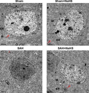
Figure 4.

Effect of NaHS on SAH‐induced neuronal ultrastructure alteration. Representative electron micrographs images showing control neurons have prominent, well‐developed, organelles: nucleus, (N), rough endoplasmatic reticulum (ER), mitochondria, (M), and ribosomes abounded (arrow). n = 4. The necrotic cells in the PFC at 48 h after SAH were fragmented, membrane dissolved and mitochondria vacuolized (asterisk *). Scale bar = 2 μm.
