
Figure 3.
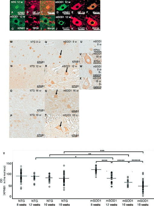
Immunohistochemistry for hypoxia‐inducible factor‐1α (HIF‐1α), karyopherin β1 (KPNB1), TAT‐interacting protein 30 kDa (TIP30), karyopherin α (KPNA) and β‐catenin in anterior horn cells (AHCs) from non‐transgenic (NTG) and mutant superoxide dismutase 1 (mSOD1) transgenic mice. Double immunostaining for KPNB1 and HIF‐1α in AHCs from 12‐week‐old NTG mice (A–C) and mSOD1 transgenic mice (D–F). Confocal microscopy of AHCs double‐immunostained for KPNB1 and HIF‐1α shows co‐localization of both proteins in the nucleus (C) and cytoplasm of AHCs (F) from 12‐week‐old NTG mice and mSOD1 transgenic mice. Confocal microscopic findings of KPNB1 and TIP30 in AHCs from 12‐week‐old mSOD1 transgenic mice (G–I). TIP30 is undetectable in AHCs (H) and is not co‐localized with KPNB1 (I). Double immunostaining for KPNB1 and nucleoporin 62 (Nup62) (J–L) in AHCs from 12‐week‐old mSOD1 transgenic mice. Confocal microscopy of AHCs double‐immunostained for KPNB1 and Nup62 shows co‐localization of both proteins on the nuclear envelope (l) of AHCs from 12‐week‐old mSOD1 transgenic mice. AHCs from NTG mice aged 8, 12, 16 and 18 weeks show nuclear immunoreactivity for KPNB1 at all examined ages (M–P), whereas nuclear immunoreactivity for KPNB1 in mSOD1 transgenic mice successively decreases as the disease progresses (allows; Q–T). The cytoplasm of AHCs from mSOD1 transgenic mice is immunopositive for KPNA (U–V) and β‐catenin (W–X), and their immunoreactivities increase as the disease progresses. The corrected OD values for KPNB1 immunoreactivity are significantly lower in mSOD1 transgenic mice aged 12, 16 and 18 weeks compared with their NTG counterparts (P < 0.05), and the value decreases significantly as the disease progresses (P < 0.05) (Y). Scale bar: 50 μm (A–K, U–X) and 100 μm (M–T). *P = 0.045; **P = 0.035; ***P = 0.0029; ****P = 0.050; *****P = 0.0029; ******P = 0.018.