
Figure 2.
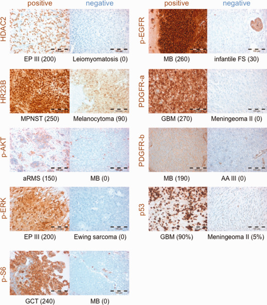
Examples of IHC staining for markers used in this study. Images depict samples with IHC stainings (brown color) scored positive (H‐Score ≥ 100) or negative (H‐Score < 100). The tumor histology is given below each image, in brackets the individual H‐Score is given, except for p53 where the percentage of positive nuclei is given. EP III: anaplastic ependymoma WHO III; MPNST: malignant peripheral nerve sheath tumor; aRMS: alveolar rhabdomyosarcoma; MB: medulloblastoma WHO IV; GCT: germ cell tumor of the pineal gland; infantile FS: infantile fibrosarcoma; GBM: glioblastoma WHO IV; AA III: anaplastic astrocytoma WHO III.