
Figure 2.
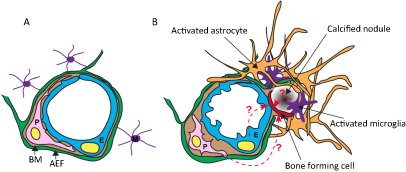
Cartoon illustrating the changes at the neurovascular unit (NVU) in idiopathic basal ganglia calcification (IBGC). The illustration takes into account observations made in genetically engineered mouse models of IBGC. A. Cross section of a normal NVU. Endothelial cells (E) are surrounded by a basement membrane (BM), pericytes (P) and astrocyte end‐feet (AEF). Microglia (M) make contacts to the NVU through cytoplasmic processes. B. Altered features of the NVU in IBGC. Calcified nodules are associated with blood vessels and are surrounded by reactive astrocytes and microglia. A hypothetical bone‐forming cell (red) initiates and propagates the formation and growth of the calcified nodule. The origin of the bone‐forming cell is unclear, but available literature provides precedence for several possible origins, as indicated (dashed line with question mark). Mouse models of IBGC provide a correlation between pericyte deficiency and BBB disruption, on the one hand, and brain calcification, on the other hand. The altered properties of endothelial cells and pericytes are indicated by their changed shape.