Figure 2.

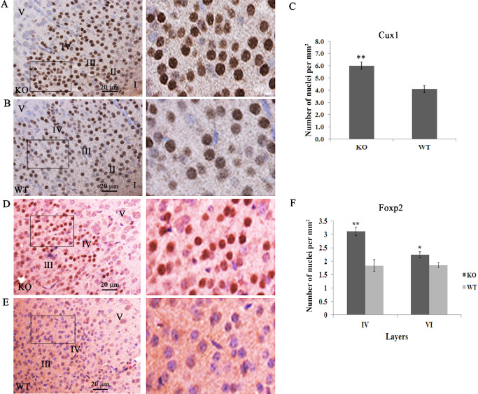
Increased cellularity in layers II–IV involves late‐born neurons. Increased cellularity of Cux1‐expressing cells in the outer cortical layers is demonstrated using Cux1‐specific antibodies in mutant null (A) compared with normal (B) mice. The boxed areas are shown at higher magnification on the right. Quantitation of Cux1‐positive cells in layers II–IV shows a ∼40% increase in the mutant mice (C). The same increase was seen for Foxp2‐positive cells in the mutant mice (D) compared with normal (E). The boxed areas are shown at higher magnification on the right. Quantitation in layers IV and VI (F) shows a significant difference in the number of Foxp2‐positive cells. (**P < 0.001; *P < 0.05).
