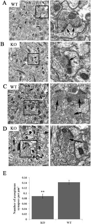
Figure 5.

Reduced spine distribution in the cortex from Lgi1 mutant mice. Electron micrographs from the cortex of P17 wild‐type (A) and mutant (KO) null mice (B) show a reduced PSD frequency (arrows) in the mutant mice. Right panels show spines in higher magnification. Analysis of vesicle‐loaded synapses (D) shows increased smaller, irregular and attenuated axonal boutons in the mutant null mice with fewer synaptic vesicles admixed with a few well‐formed vesicle‐rich asymmetric synapses (right) compared with mostly well‐formed axonal boutons in P17 normal mice (C, shown at higher magnification on the right). Quantification of functional synapses (see text) demonstrates a ∼35% reduction in the mutant mice (E) (**P < 0.001).
