
Figure 5.
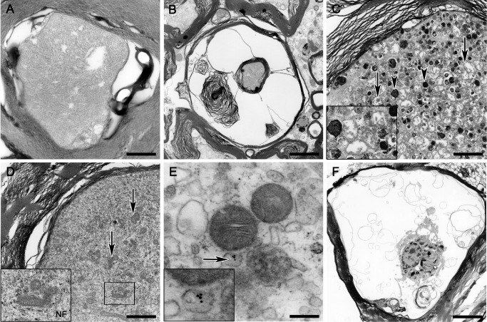
Transmission electron microscopy. (A) Intact axon of a control animal exhibiting mild myelin changes interpreted as fixation artifacts. (B) Myelin sheath dilatation and myelin fragmentation in an animal with acute SCI. (C) Axon swelling and axoplasmic degeneration with accumulation of numerous electron‐dense bodies (arrowheads; inset) and swollen mitochondria (arrows; inset) in an animal with subacute SCI. (D) Marked axoplasmic accumulation of mitochondria (arrows; inset) intermingled with numerous neurofilaments (inset) without dense body formation in a swollen axon of an animal with subacute SCI. (E) Immunoelectron microscopic illustration of potentially microtubule‐associated gold particles specifically labeling growth‐associated protein‐43 (arrow, inset) in a mitochondria‐rich swollen axon of the same animal as in (D). (F) Large phagocytic cell located within an empty myelin sheath showing phagocytosed myelin and axonal remnants in an animal with subacute SCI. A scale bar = 0.5 μm; B scale bar = 2.7 μm; C,D scale bar = 1.7 μm; E scale bar = 0.2 μm; F scale bar = 5.4 μm. SCI = spinal cord injury.