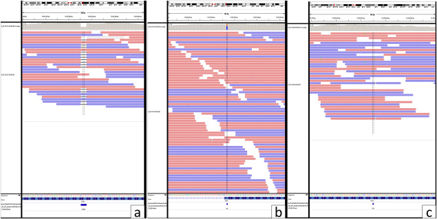
Figure 3.

Representative photographs showing ATRX ( A,B ) and DAXX ( C ) mutations Red reads mapped to forward strand and Blue mapped to reverse strand. Bases that differ from the reference are displayed with their letters in color. A. ATRX mutation—Frameshift variant ENSP00000362441.4:p.Lys1045Ter. B. ATRX mutation‐ Stop gain‐ and splice region variant ENSP00000362441.4:p.Ser1245Ter. C. DAXX mutation—Missense variant NSP00000363668.5:p.Asp331Asn.
