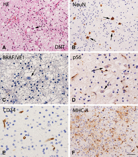
Figure 3.

Dysembryoplastic neuroepithelial tumor (DNT; BRAF wild‐type status): histopathological features, VE1 and pS6, CD34 and MHC‐II immunoreactivity. A. Hematoxylin & eosin (HE) staining of DNT showing a typical heterogeneous cellular composition, with floating neurons (arrow) surrounded by a prominent population of oligodendroglia‐like cells. B. NeuN staining detects the neuronal component of DNT. C. No detectable BRAF V600E (VE1) immunoreactivity (IR) within the tumor area. D. No detectable phosphorylated (p)‐S6 IR in neuronal cells (arrows). E. CD34 shows IR only in blood vessels. F. MHC class II antigen (MHC‐II) expression within the tumor area (microglial cells). Scale bar in (F): A, B, F: 80 μm; C, D, E: 40 μm.
