
Figure 3.
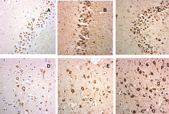
FUS pathology in aFTLD‐U (A–C) and Neuronal Intermediate Filament Inclusion Body Disease (D–F), as shown by immunostaining for FUS protein (A,D), transportin‐1 (B,E) and TAF15 (C,E).
Official websites use .gov
A
.gov website belongs to an official
government organization in the United States.
Secure .gov websites use HTTPS
A lock (
) or https:// means you've safely
connected to the .gov website. Share sensitive
information only on official, secure websites.
FUS pathology in aFTLD‐U (A–C) and Neuronal Intermediate Filament Inclusion Body Disease (D–F), as shown by immunostaining for FUS protein (A,D), transportin‐1 (B,E) and TAF15 (C,E).