
Figure 1.
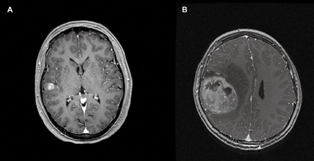
A. Post‐gadolinium t1 axial magnetic resonance image demonstrating a well‐defined enhancing superficial ePXA in the right temporal lobe, involving mostly the cortex; surrounding edema is not seen. B. Post‐gadolinium magnetic resonance t1 axial image demonstrating a well‐defined, heterogeneously enhancing superficial eGBM with marked surrounding edema.