
Figure 2.
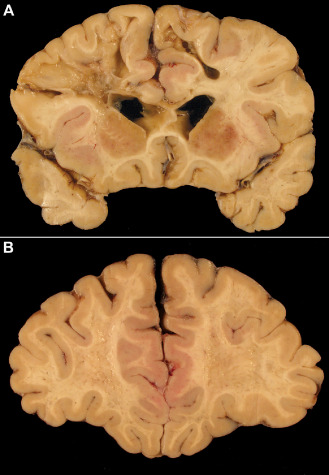
Ischemic white matter damage in a dementia patient with cardiac valvular disease and recurrent episodes of bradyarrhythmia. A. A coronal slice through the frontal and temporal lobes reveals multiple cavitated infarcts and foci of softening and gray discoloration. There is only mild atherosclerosis. B. In a more anterior slice through the same brain, the white matter appears pitted, with numerous confluent foci of gray discoloration.