
Figure 5.
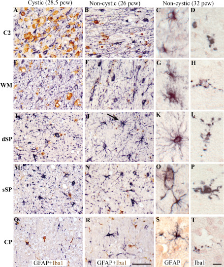
In vertical columns: different cases of periventricular white matter injury (PWMI): a cystic case at 28.5 pcw (A,E,I,M,Q), a non‐cystic case at 26 pcw (B,F,J,N,R) and a non‐cystic case at 32 pcw (C,D,G,H,K,L,O,P,S,T). Immunolabeling for glial markers from deep to superficial cerebral wall layers: area C2 (A–D), superficial white matter (WM) (E–H), deep subplate (dSP) (I–L), superficial subplate (sSP) (M–P), cortical plate (CP) (Q–T). Double‐labeling of glial fibrillary acidic protein (GFAP)‐positive astrocytes (black) + ionized calcium‐binding adapter molecule 1 (Iba1)‐positive microglia/macrophages (brown) (A,D,G,J,M and B,E,H,K,N). GFAP‐positive astrocytes (C,G,K,Q,S) and Iba1‐positive microglia/macrophages at higher magnifications (C,F,I,L,O and D,H,L,P,T). The arrow in J shows a big protoplasmic astrocyte with long processes located in the dSP in a non‐cystic lesion. Scale bars: A,B,E,F,I,J,M,N,Q,R. 50 μm; C,D,G,H,K,L,O,P,S,T. 15 μm.