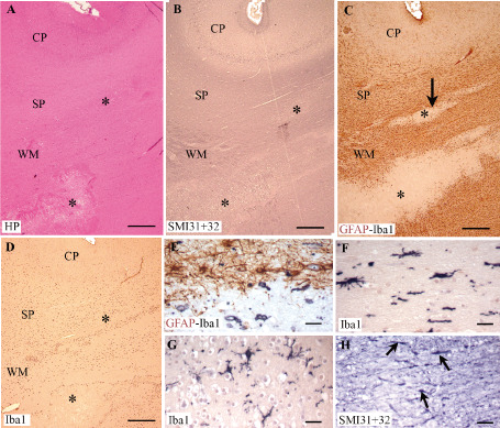
Figure 7.

Serial sections of a non‐cystic lesion in the cerebral wall of a preterm infant (34.5 pcw) showing necrotic foci in the white matter (WM) and subplate (SP) (asterisks). A. Hemalum‐phloxine (HP) staining. B,H. SMI31 + 32 immunolabeling. C,E. Double‐labeling for GFAP (brown) + ionized calcium‐binding adapter molecule 1 (Iba1) (black). D,F,G. Iba1‐positive microglia/macrophages. The arrow in C indicates the lesion in the SP, enlarged in E and H. Note the Iba1‐positive macrophage in the deep SP in E, whereas Iba1‐positive microglia have an intermediate phenotype in the superficial SP (G) and cortical plate (CP) (F). In H, note the SMI‐positive axonal spheroids (small arrows) present in the deep SP. Scale bars: A,B,C,D. 500 μm; E,F,G,H. 50 μm.
