
Figure 8.
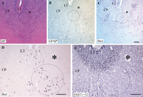
Serial sections of a non‐cystic lesion in the cortical plate (CP) of a preterm infant (33.5 pcw). Hemalum‐phloxine (HP) staining of a lesion located in the CP (A). Patches of cells positive for glial fibrillary acidic protein (GFAP) (B) and ionized calcium‐binding adapter molecule 1 (Iba1) (C,D) in the same area delineated by a circle. The asterisk show the same vessel visualized on the different serial sections in A–E. The circle in C is enlarged in D. In E, note the spot with no SMI31 + 32‐positive neuronal labeling within the circle at the level where numerous Iba1 macrophages are present in D. L1, layer 1. Scale bars: A–C. 200 μm; D,E. 100 μm.