
Figure 5.
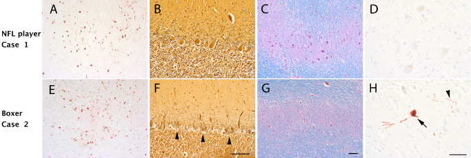
Neuropathology and cerebellar degeneration in a former professional boxer and football player. Case 1 (A–D): A 56‐year‐old former National Football League (NFL) player had chronic traumatic encephalopathy (CTE) characterized by phosphorylated tau‐positive neurofibrillary tangles present at the sulcal depths (A, AT8 immunostain). His cerebellum appeared intact with a well‐populated Purkinje cell layer (B, Bielschowsky silver stain) and dentate nucleus (C, Luxol hematoxylin and eosin). There was no phosphorylated tau accumulation present within the dentate nucleus (D). Case 2 (E–H): A 61‐year‐old former professional boxer had a similar degree of CTE‐related tauopathy (E, AT8 immunostain). In fact, both the NFL player and the boxer met neuropathological criteria 49 for CTE stage III tauopathy. However, in contrast to the NFL player, there is marked degeneration within the cerebellum with marked Purkinje cell loss (F, Bielschowsky silver stain; arrowhead, Basket cell processes without Purkinje cells “empty Baskets”). There is also loss of neurons within the dentate nucleus (G), which contains scattered phosphorylated tau‐positive neurons (H, arrow) and processes (H, arrowhead). These two cases illustrate the need for careful clinical phenotyping and demonstrate the utility of recently published criteria 61. Scale bars, A, C, E and G, 100 μm; B and F, 100 μm; D and H, 50 μm.