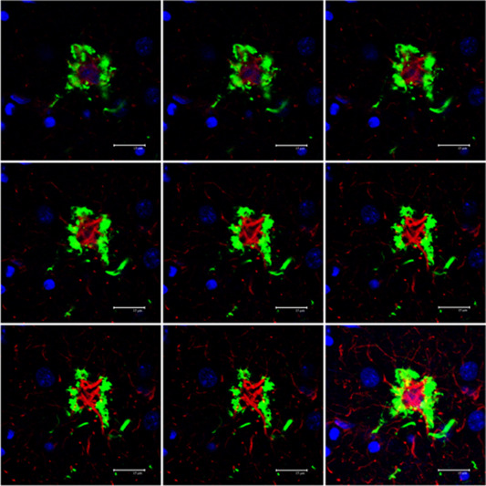
Figure 5.

Serial reconstruction of a tufted astrocyte in PSP processed for double‐labeling immunofluorescence and confocal microscopy using antibodies to P‐tau (AT8, green) and GFAP (red). Note tufts of P‐tau at the periphery of the cytoplasm and proximal part of astrocytic branches, and redistribution of GFAP at the inner part of the cytoplasm with very poor GFAP immunostaining of astrocytic branches. Nuclei (blue) are stained with DRAQ5TM; bar = 15 μm.
