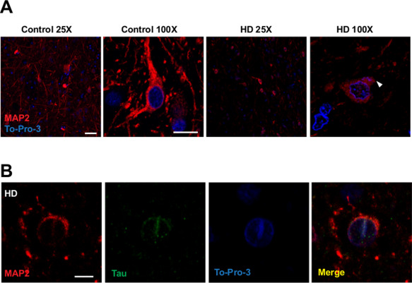
Figure 5.

Dendritic loss of MAP2 in striatal HD neurons and detection of MAP2 nuclear deposits in striatal neurons. A. Images show MAP2 immunofluorescence (red) with a To‐Pro‐3 counterstaining (blue) of striatum from controls and HD patients. Images are a single stack representative of all cases analyzed n = 4/5. Loss of MAP2 in dendritic arbors in HD neurons. Scale bar 50 µm in 25X images, 7,5 µm in 100X images. White arrowhead shows MAP2 stacked at the base of the dendrite in striatal HD neurons. B. MAP2 and MAP Tau nuclear deposit in HD striatal neuron. Scale bar 5 µm.
