
Figure 4.
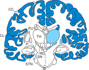
Distribution and severity of neuronal loss in select brain regions of Huntington's disease (HD) patients. Schematized frontal section through the cerebral hemispheres at the level of the red nucleus (RD) (brain regions undergoing severe neuronal loss are highlighted with blue shading and brain regions with marked loss of nerve cells with lighter blue shading). (Abbreviations: C, caudate nucleus; CC, cerebral cortex; CA, corpus callosum; CL, claustrum; HD, Huntington's disease; P, pallidum; PU, putamen; RD, red nucleus; SN, substantia nigra; STN, subthalamic nucleus; TH, thalamus).