Figure 2.

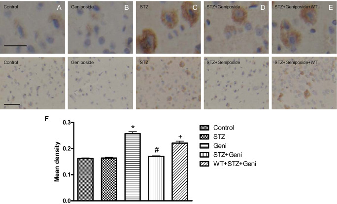
Photomicrographs and quantitative analysis of immunohistochemistry of phospho‐tau in the rat cerebral cortex. Immunoreactivity of phospho‐tau was presented as brown staining detected under higher and lower magnification, respectively (A–E, above bar = 50 μm; below bar = 150 μm). STZ treatment significantly increased phospho‐tau compared with controls (A,C,F; *, compared with control, P < 0.01), but geniposide (Geni) treatment prevented STZ‐induced increase in phospho‐tau (C,D,F; #, compared with STZ group, P < 0.01); even injection of geniposide alone did not induced the remarkable change of phosphor‐tau (A,B). PI3K inhibitor wortmannin (WT) partially prevented the protective effect of geniposide (E,F; +, compared with STZ + Geniposide group, P < 0.01).
