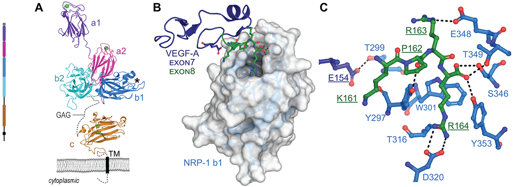
Figure 1. Schematics of NRP-1 domains, VEGF-A165 binding, and CendR interaction network.

A. Domain architecture of NRP-1 with domain a1 from mouse (PDB ID 4GZ9)94, domains a2, b1, b2 (PDB ID 2QQM)10 and MAM (PDB ID 5L73)11 from human. Bound Ca2+ shown as green spheres, missing loops as dashes, transmembrane domain as a rectangle. GAG indicates region of glycosaminoglycan modification. *Indicates VEGF-A interaction pocket. B. Structure of NRP-1 b1 domain, shown as white surface, in complex with the heparin binding domain (exon 7/8) of VEGF-A164, shown as cartoon with exon 7 in dark blue, exon 8 in green (PDB ID 4DEQ)15 C. Details of VEGF-A Glu154 and KPRR164 interactions with NRP-1 (close up of view B). Dashes indicate polar or salt-bridge contacts within 3.0 Å.
