
Figure 1.
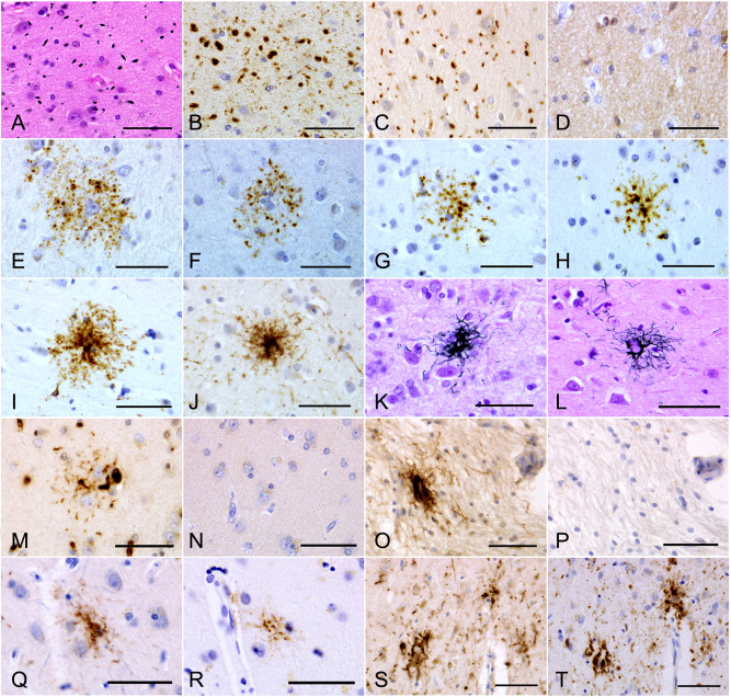
A–D. Argyrophilic grains in the amygdala in an AGD‐TA case. Argyrophilic grains are (A) Gallyas‐positive, (B) AT8‐positive, (C) RD4‐positive, but (D) RD3‐negative. E–G. AT8‐positive TAIs in the frontal cortex in AGD‐TA cases lacking Gallyas‐positive TAs in any region. Fine dot‐like granular phosphorylated tau accumulations surround the nuclei of astrocytes. H, I. AT8‐positive astrocytic lesions in the frontal cortex in AGD‐TA cases having Gallyas‐positive TAs. Phosphorylated tau tends to be densely accumulated around the nuclei of astrocytes. J. An AT8‐positive astrocytic lesion in a PSP case. Fibrous rather than dot‐like structures are densely distributed in the central portion. Their morphological features tended to be similar to those in AGD‐TA cases having Gallyas‐positive TAs (H, I) rather than in AGD‐TA cases lacking them (E, F, G). K, L. Gallyas‐positive TAs in the frontal cortex in (K) AGD‐TA and (L) PSP cases. They could not be distinguished morphologically. M, N. (M) An RD4‐positive TAI in an AGD‐TA case. (N) The lesion lacked RD3 immunoreactivity. Serial sections from the caudate nucleus. This case lacked Gallyas‐positive structures in this region. O, P. (O) An RD4‐positive astrocytic lesion in a PSP case. (P) The lesion lacked RD3 immunoreactivity. Serial sections from the putamen. Q, R. (Q) An RD4‐positive astrocytic lesion in the frontal cortex in an AGD‐TA case having Gallyas‐positive TAs. (R) The lesion also showed AT8 immunoreactivity. The identical region in mirror serial sections is shown after reversal. S, T. (S) RD4‐positive astrocytic lesions in the frontal cortex in a PSP case. S. The lesions also showed AT8 immunoreactivity. T. Mirror serial section of the identical region. All scale bars = 50 μm.