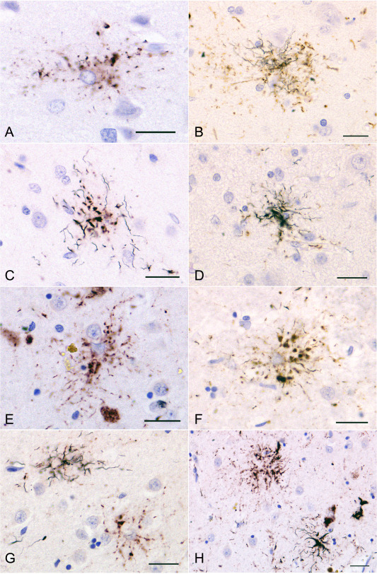
Figure 4.

Double staining of AT8 immunohistochemistry and Gallyas method in an AGD‐TA case having Gallyas‐positive TAs and a PSP case. A, B, C, D. TAIs (A) and TAs (B, C, D) in the superior frontal cortex in an AGD‐TA case (AGD‐TA9 in Table 4). The proportion of Gallyas‐positive structures varied between lesions. In some TAs, fine argyrophilic thread‐like structures were found only in the distal portion of each astrocytic lesion (C). E, F, G, H. TAIs and TAs in the putamen in a PSP case. (E) As in AGD‐TA cases, TAIs had fine granular tau accumulations that were radially arranged. It is hard to morphologically distinguish TAIs in PSP cases (E) from TAIs in AGD‐TA cases (A). F. When the number of Gallyas‐positive thread‐like structures was small, they were often found in the distal portion of each astrocytic lesion. G. The lesion on the right lacks a Gallyas‐positive structure (ie, TAI), while the one on the left has Gallyas‐positive thread‐like structures (ie, TAs). H. A right‐hand lesion has Gallyas‐positive thread‐like structures (ie, TA), while the left‐hand one lacks Gallyas‐positive structures (ie, TAIs). The peroxidase labeling was visualized with DAB (B, D, F) or Vector NovaRED (A, C, E, G, H). All scale bars = 20 μm.
