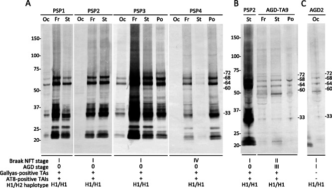
Figure 9.

Immunoblot analysis of the sarkosyl‐insoluble, urea‐soluble fraction with T46 from frozen brain tissues of PSP A, B, AGD‐TA B and AGD C cases. A. Approximately 68‐ and 64‐kDa bands, as well as the 33‐kDa low‐molecular mass tau fragments characteristic of PSP, are seen in the frontal cortex, striatum, pons and/or occipital cortex in all PSP cases. In addition, a minor 60‐kDa band that may reflect the coexistence of AD pathology is seen in some lanes in PSP1, PSP3 and PSP4. B. Both 68‐ and 64‐kDa bands, as well as the 33‐kDa low‐molecular mass tau fragments characteristic of PSP, are found in not only a PSP case (PSP2) but also an AGD‐TA (AGD‐TA9) case having Gallyas‐positive TAs. C. Both 68‐ and 64‐kDa bands are seen in an AGD case. Although this case did not have any TAs or TAIs, a minor 33‐kDa band is also observed. Genetic analysis demonstrated that all cases had the H1/H1 haplotype. Fr = frontal cortex; St = striatum; Po = pons; Oc = occipital cortex; TAs = tufted astrocytes; TAIs = tufted astrocyte‐like astrocytic inclusions.
