
Figure 1.
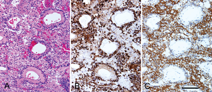
Serial sections from case 2 show a pseudopapillary architecture (A) with hyalinized blood vessels surrounded by a layer of GFAP‐immunoreactive glial cells (B). The interpapillary areas are occupied by sheets of synaptophysin‐immunoreactive neurocytic and ganglioid cells mixed with GFAP‐positive glial cells. (A) H&E, (B) GFAP immunostain, (C) synaptophysin immunostain. Bar = 100 microns.