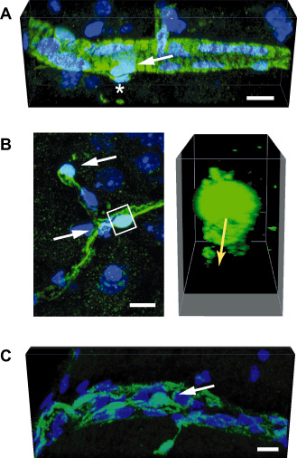
Figure 2.

Mural cells of the central nervous system (CNS) vasculature. This figure demonstrates the different phenotypes of mural cells along different segments of the CNS vasculature. The mural cells can be identified based on their expression of the green fluorescent protein (GFP) in a mouse line where the pericyte‐specific rgs5 gene has been replaced with a GFP reporter 56. The images represent maximal projections or three‐dimensional (3D) renderings of confocal stacks obtained from GFP‐expressing cells in the brains of rgs5 GFP/ GFP mutant mice. Nuclei are counterstained with 4′,6‐diamidino‐2‐phenylindole (DAPI). Scale bars: 10 μm. A. In a precapillary arteriole, mural cells (arrow) exhibit robust and densely packed circumferential processes, suggesting a contractile phenotype. An abluminal GFP‐expressing cell is marked by an asterisk. B. Pericytes investing capillaries (white arrows in the left panel) show a typical protruding fusiform cell body with long primary processes running in the length of the capillary. The primary processes give rise to perpendicular secondary processes. The 3D reconstruction (right panel) of the volume marked by the white box demonstrates circumferential processes underneath the pericyte cell body that surround the capillary lumen (indicated by the yellow arrow). C. Mural cells (arrow) in a postcapillary venule show a flattened, stellate morphology and loosely cover the length of the vessel.
