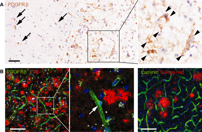
Figure 4.

Fibrosis in human neurological disorders. Conventional and laser confocal microscopic images of immunostained human post‐mortem brain sections. Nuclei are counterstained with hematoxylin or 4′,6‐diamidino‐2‐phenylindole (DAPI). Scale bars: 100 μm. A. Platelet‐derived growth factor receptor (PDGFR)‐β‐immunoreactive cells (brown) in a subacute stroke lesion. PDGFR‐β‐immunoreactive cells are associated with vessels in the non‐ischemic tissue (arrows), but detach from the vasculature and spread into the parenchyma (arrowheads) in the ischemic tissue (right of the white dashed line). B. PDGFR‐β‐immunoreactive cells in Alzheimer's disease (left and middle panels). PDGFR‐β‐immunoreactive cells (green) remain at perivascular sites. There is no apparent proliferation of PDGFR‐β‐immunoreactive cells around amyloid plaques (red). In addition, no ectopic deposition of laminin (green) can be detected around plaques (red) in the Alzheimer's disease brain (right panel).
