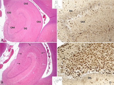
Figure 4.

The left pictures show the physiological ( A ) and pathological ( B ) subgross appearance of the temporal hippocampus. B features the most common polysegmental HS with losses throughout all CA segments (black arrows) and a few residual pyramidal clusters (white arrows) in CA1. On the right, GFAP‐positive astrocytes are shown in the unaffected hilus (C) and this of a cat with severe HS (D).
