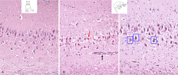
Figure 5.

(A) Pyramidal cell degeneration in limbic encephalitis compared to a normal CA segment. (B) Neuronal loss shows morphologic features of excitotoxicity in terms of eosinophilic necrosis (red arrow). The black arrow indicates scanty perivascular inflammation. (C) Note the perineuronal infiltrates (blue frames).
