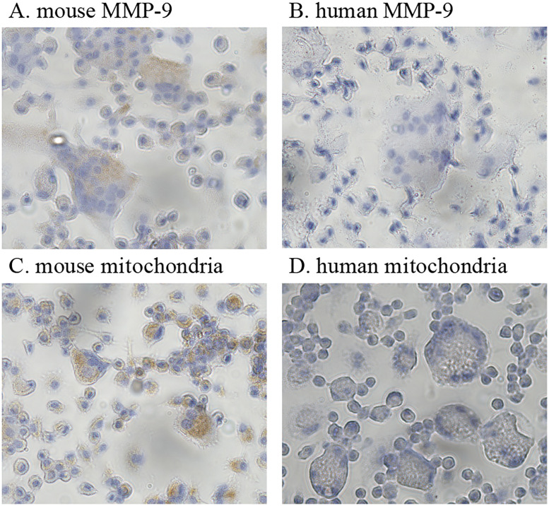
Fig 5. Cytochemistry of cultured mouse osteoclast progenitor cells in the presence of M-CSF and RANKL differentiation signals.
(A–D) Cultured cells from mouse osteoclast progenitor cells. (A) Cultured cells immunostained with an antibody that reacts with mouse MMP-9. Multinucleated cells positive for MMP-9 stained brown with the anti-MMP-9 antibody (dilution, 1:100). (B) Cultured cells immunostained with an antibody specific to the human MMP-9. Multinucleated cells were not stained (antibody dilution, 1:12.5). (C) Cultured cells immunostained with an antibody that reacts with mouse mitochondrial protein. Multinucleated cells positive for the mouse mitochondrial protein stained brown with the anti-mitochondrial antibody (dilution, 1:360). (D) Cultured cells immunostained with an antibody specific to human mitochondrial protein. Multinucleated cells were not stained (antibody dilution, 1:10). Original magnification, 400×.

