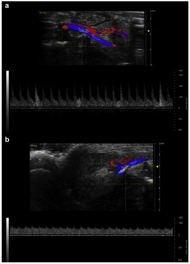
Figure 2 ∣. Cecal ligation and puncture (CLP) alters renal arterial pulsewave Doppler waveform morphology.
Representative images of pulsewave Doppler recording used to measure renal arterial velocities. Blue indicates flow away from the ultrasound probe (toward the kidney) and red toward the probe (away from the kidney). Waveforms show arterial velocity on the Y-axis and time on the X-axis. (a) T0 mouse. (b) Mouse 48 hours after CLP.

