Figure 6:

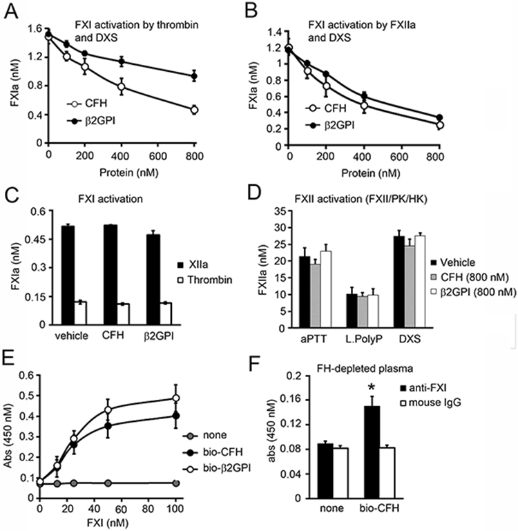
Inhibition of FXI activation by CFH. (A) FXI (30 nM) was incubated with either thrombin (5 nM) or (B) FXIIa (5 nM) in the presence of dextran sulfate (DXS, 2 ug/ml) for 15 min at 37°C in the presence or absence of increasing concentrations of factor H (0-800 nM) or β2GPI. FXIa generation was measured with a chromogenic substrate. (C) FXI (100 nM) was incubated with either thrombin (10 nM) or FXIIa (10 nM) in the absence of DXS for 30 min at 37°C in the presence or absence of factor H (800 nM) or β2GPI (800 nM). FXIa generation was measured with a chromogenic substrate. (D) FXII activation was measured following the addition of 200 nM FXII, 50 nM PK and 50 nM HK in the presence of aPTT reagent, 10 μM long polyP or DXS (2 μg/ml) in the presence or absence of factor H or β2GPI (800 nM). FXIIa generation was measured with a chromogenic substrate. (E) 96-well plates were coated with 5 μg/ml streptavidin prior to addition of vehicle, biotinylated CFH (bio-CFH) or β2GPI (bio- β2GPI) (100 nM). Increasing concentrations of FXI was added and binding was detected with a specific antibody against FXI. (F) 96 well plates were coated with 5 μg/ml streptavidin prior to addition of vehicle or biotinylated CFH (bio-CFH). FH-depleted plasma was added and binding was detected with a specific antibody against FXI or a mouse IgG control. * P < 0.05 with respect to vehicle. Mann-Whitney U test was used for statistical comparisons. Data are mean ± SD (n = 3).
