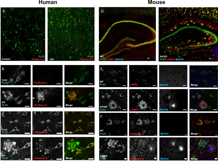
Fig 1. PLD3 is enriched in lysosomes surrounding parenchymal β-amyloid plaques in AD tissue.
A. Low magnification images of temporal lobe cortex from neurological control and AD brain demonstrated a neuronal pattern of staining against PLD3 (green) and consistent accumulation around β-amyloid plaques. B. 40x magnification images of the same immunohistochemistry show PLD3 is in a punctate staining pattern within neuronal cell bodies and enriched around β-amyloid plaques. C. Co-staining with lysosomal marker cathepsin B confirmed PLD3 was primarily lysosomal and enriched in dystrophic neurites in AD brain. D. In 5xFAD mice, PLD3 was similarly enriched around every β-amyloid plaque and strongly colocalized with LAMP2. E. 40x magnification of WT and 5xFAD tissue demonstrated strong co-localization of PLD3 with lysosomal membrane marker LAMP2 (Pearson coefficient: 0.84±0.07 for WT, 0.86±0.04 for 5xFAD) and strong staining in dystrophic neurites around β-amyloid plaques, which are stained blue with methoxy-X04. F. The lysosomal lumenal protease cathepsin B is similarly co-localized with PLD3 in both normal and diseased neurons.

