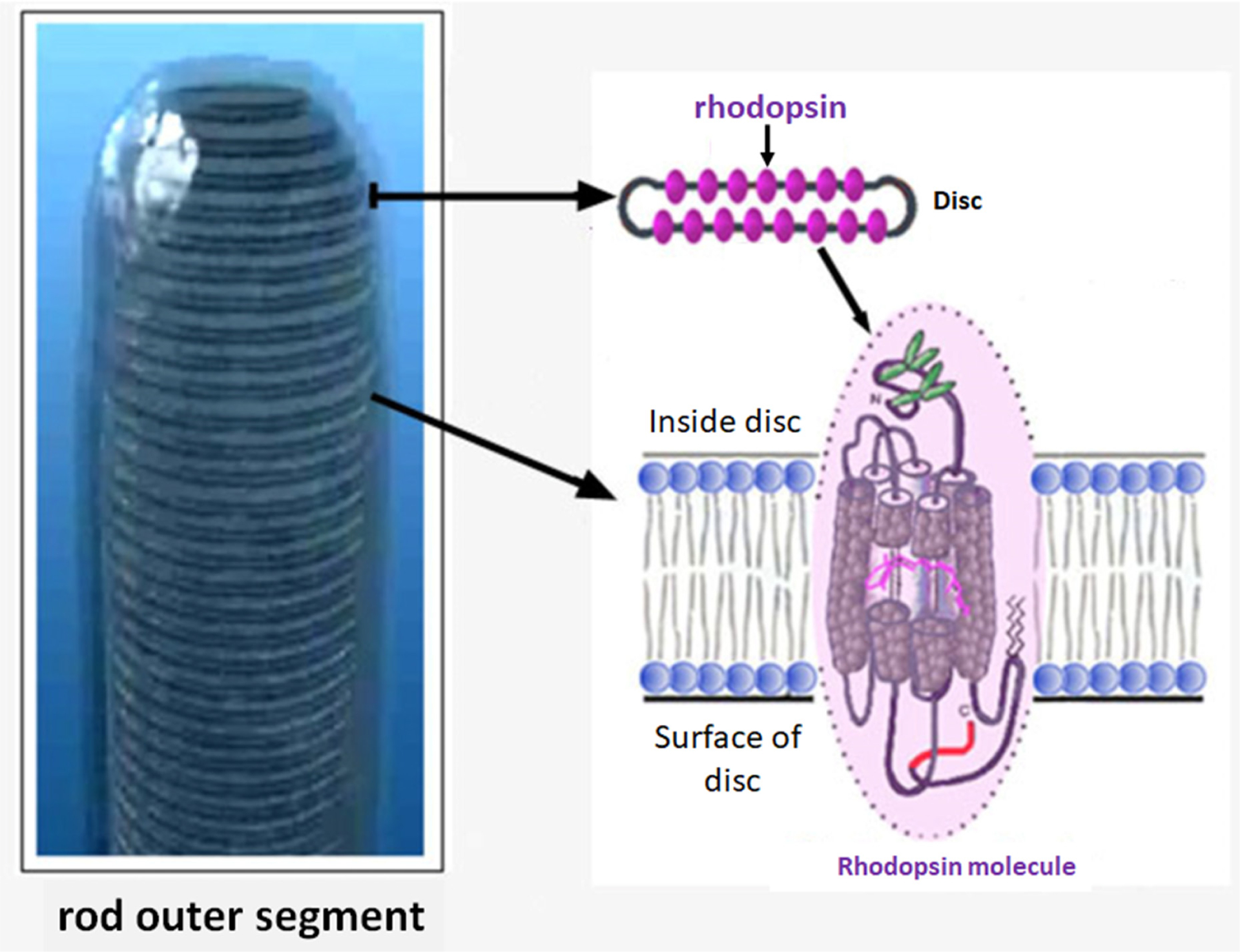
Figure 2.

Structure of the rod outer segment with densely packed discs, each expressing thousands of rhodopsin molecules. Note that the stacked discs are contained within the plasma membrane of rod outer segment. Each stacked disc contains multiple rhodopsin molecules oriented with the carboxyl terminus facing the cytoplasm (surface of the disc) where GRK1, Gt1 and arrestin are located, as well as other regulators of visual signalling. 11-cis-retinal is shown within the transmembrane domains of rhodopsin. Adapted from Webvision, webvision.med.utah.edu/. Noncommercial 4.0 International (CC BY-NC) Creative Commons license.
