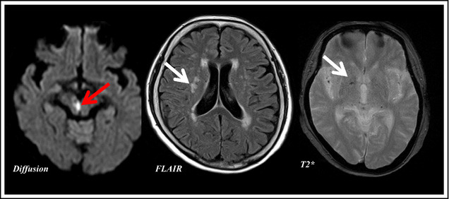
Figure 1.

Imaging of a 72‐year‐old woman who developed a morning‐onset stroke. The paramedian branch of the posterior cerebral artery exhibited signs of branch atheromatous disease, and this anatomical location corresponded to the site of high intensity of diffusion magnetic resonance imaging and her neurological deficit. The red arrow indicates the acute infarction in diffusion magnetic resonance imaging and the white arrows show old deep white matter infarcts (fluid‐attenuated inversion recovery) and microbleed (T2*).
